Making an artificial immune niche work
Written by: Janet Huisman, Øyvind Halaas and Otto Paans
In the INCITE project we are building an artificial immune niche: an academy for T cells where they learn the best ways to attack cancer cells. But to make the academy function smoothly, we need to focus on three main components: the cells, the scaffold and the fluid flow.
The cells are much like an academic community. We can divide them into three groups:
- The T cells are the students that need to learn how to recognize and fight the cancer.
- The dendritic cells are their teachers, who have studied the cancer cells and know what they look like and how to neutralize them.
- The fibroblasts (or stromal cells) are supporting personnel, who provide and maintain the conducive environment that is required for a good education. They do this for example by supplying certain molecules and creating a surface for the other cells to attach to.
The scaffold comparable to the academy building, which provides the cells a three-dimensional space to live and learn inside. It is important that this academy building contributes to the health of the inhabiting cells so that they thrive inside. We can construct the scaffold in almost any shape we want, thanks to the 3D printing technology of our project partner UpNano. This is important, as finding the right shape and size for the scaffold is a design process – we need to experiment, try new options and find the optimal solution.
Within the scaffold, we also require a fluid flow. Think of it as a fresh supply of air and a good catering service in the academy. While we humans need oxygen in the air, the cells in your body are actually more like fish and they like to live in water. (This is why more than 60% of your body is made up of water). And just like fishes in an aquarium, cells need a fresh supply of water from time to time to provide them with food and oxygen and to remove their waste.
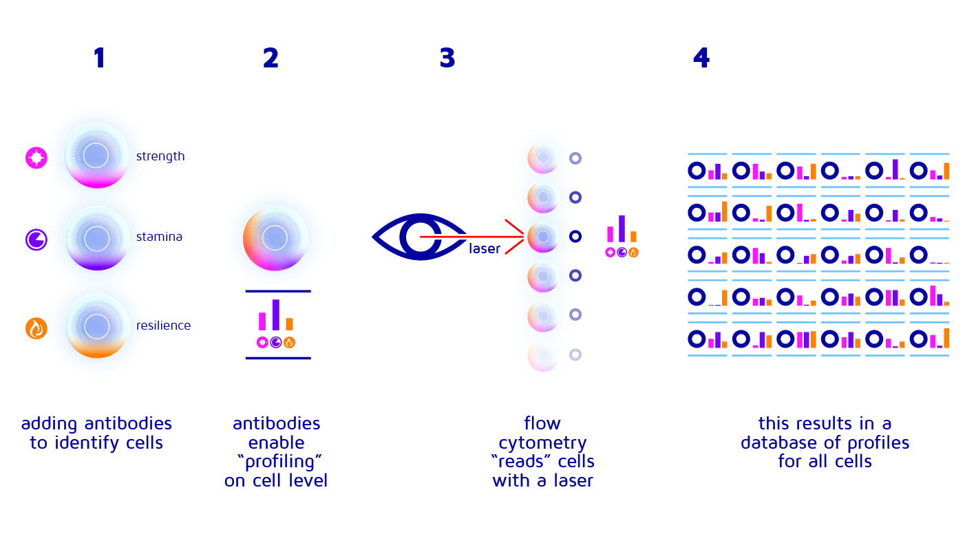
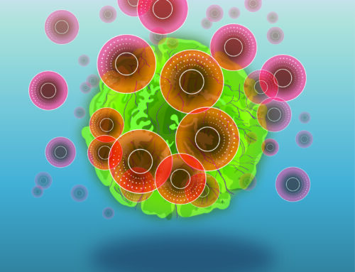
Figure 1: Method of flow cytometry. Antibodies added to cells allow them to be profiled and read. This allows for building a performance profiles that can be used to select the best specimens
Since we know which colour represents which characteristic, we then know a lot more about our cells and which characteristics they have. Basically, we colour-code cells, giving each of them a coloured “barcode” or “performance profile” that we can read and interpret to get a good overview of the cell collection.
But only because things may look right doesn’t mean they work correctly. So, we must check T cells “in action” too. While we may be able to confirm whether a certain type of T cell has a good memory, or is really strong, we still cannot be certain how it will perform when actually fighting cancer cells. A cell that has all the right characteristics may still not be an optimal “soldier”.
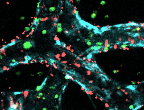
2D/3D cancer tests
Third, instead of only looking at interactions and characteristics, we also look at their effectiveness in killing cancer cells. These so-called anti-cancer killing assays, or cytotoxicity assays, is the third way of analysing the cells in our project. The easiest way is to check if they produce certain molecules that indicate a high cancer-killing activity. Even better would be to add the T cells that we have been teaching in our academy to a dish with some cancer cells and see how many they can kill. But the only way to really be sure that these T cells are strong enough is to inject them back into the mouse where they came from and observe whether they can kill the entire tumour. This is the ultimate preclinical lab test of their capabilities and serves to select only the best protocols for making cells based on their characteristics and performance. If at some later point in time, we are ready to see whether this works also for human patients in the real world, there are strict regulations and large-scale documentation that must be completed in many cancer centres for many years. So, we better have made the correct choices early on to prevent costly failures later on!
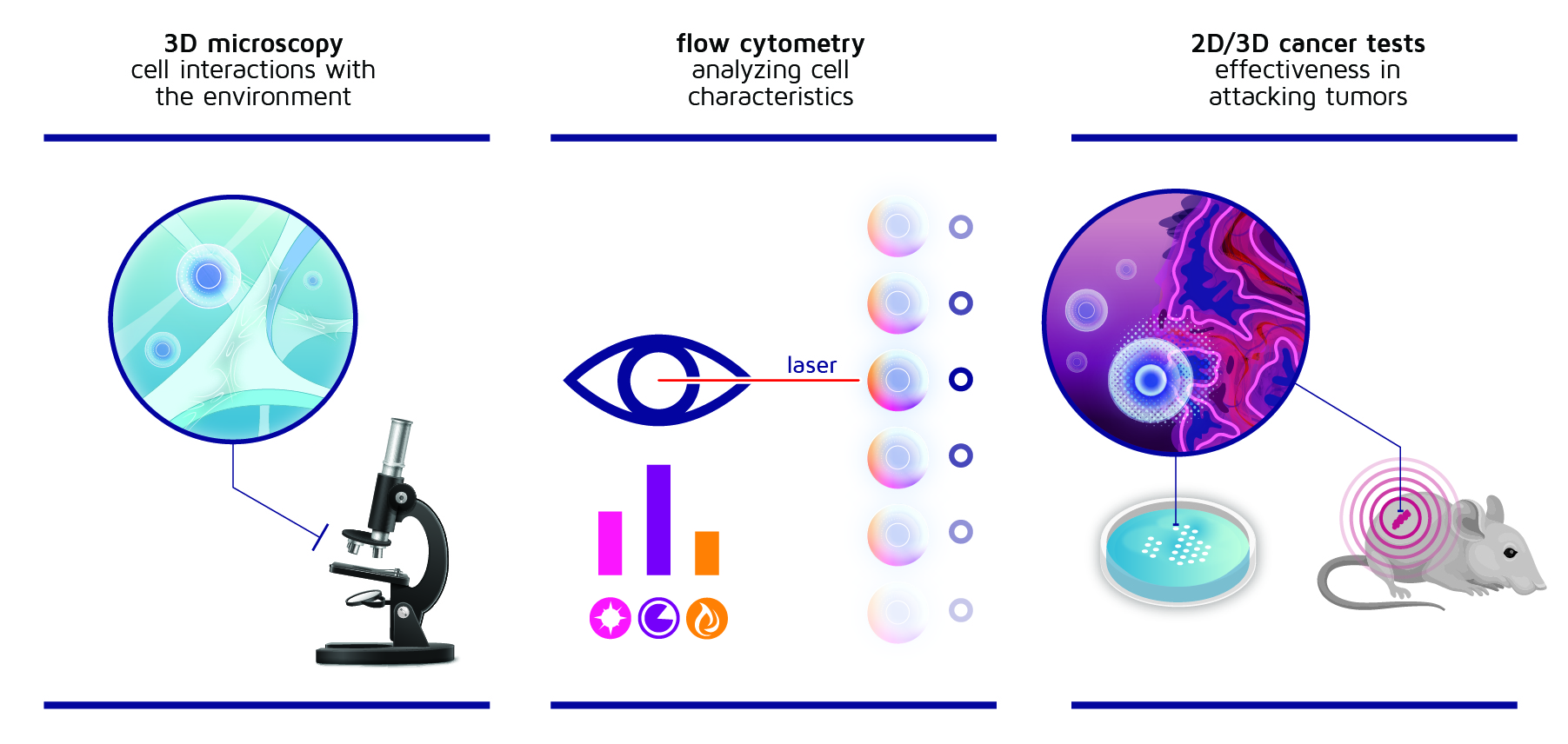
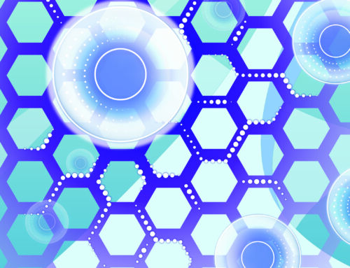
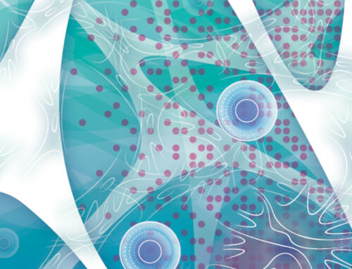
Figure 2: Three of the four assessment methods used in the INCITE project
Now that we know which components we need, of course we could just combine everything and hope it will work on the first try. But if it doesn’t (which is very likely), how can we find out what to change? To address this issue, we start with a simple setup and slowly increase the level of complexity, so that we understand exactly what is going on in each step and we can point out where things are going wrong. By building step-wise from simple to complex, we can easily see where we took a wrong turn, or where things did not work out as we envisioned.
The first experiments we attempted were therefore not very complex. We just added three different cell types in a dish and observed what happened. Sounds easy, right?
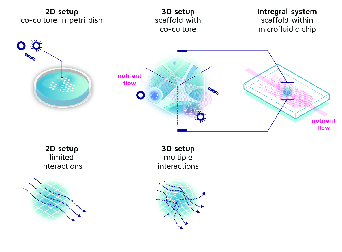
Figure 3: From simple petri dish and co-culture to an increasingly complex setup with cells in a 3D scaffold. The scaffold is located in a microfluidic chip that provides a steady nutrient flow.
In the INCITE project, these three assessment methods (3D microscopy, flow cytometry, and 2D/3D cancer tests) are used to understand the functionality of the artificial immune niche and to learn how to improve it. With every experiment we learn something new about how many cells we need, which scaffold design works best, and which fluid flow rate provides the best education for the T cells. Consequently, we learn much about the characteristics that the T cells need to have to become effective killers. And we really need all of this knowledge to make sure that the academy is teaching the T cells in the best possible way how to fight cancer.