Modeling the Efficacy of T Cells
Written by: Pierre van den Brugge, Luca Gattinoni and Otto Paans
In the INCITE project, we are designing and constructing an artificial immune niche—akin to a military academy. Here, T cells are trained like future soldiers, becoming skilled to recognize and effectively attack cancer cells, and to survive in harsh environments.
Our program is highly specific: T cells are taught to recognize tumor cells, each marked by a unique “antigen” that identifies it as “the enemy.” In this educational program, T cells learn to attack upon recognizing this mark, and we further enhance their ability by using CAR-T cells—immune cells that are modified with a special receptor to specifically target and destroy cancer cells. To test them, we see how they respond in various environments, in this case, in vitro (cell-based) and an in vivo (organism-based) settings.
In vitro testing
After the “T cell soldiers” have completed their training, we want to ensure they are fully prepared for operating in the field. Instead of sending them directly to battle and see what happens, we will check beforehand if they possess the requisite characteristics of good soldiers. Using flow cytometry (explained in blog 7), we assess the presence of certain surface markers on T cells when they exit the artificial immune niche, confirming their “memory stem cell” features. Like a soldier with armor, T-cells with such a differentiation state have enhanced survival and proliferation when injected into patients.
We ensure T cells are well-equipped with the right weapons. Through various techniques, such as ELISA, we verify their ability to produce molecules necessary to kill cancer cells (see blog 7 for further details on the verification process).
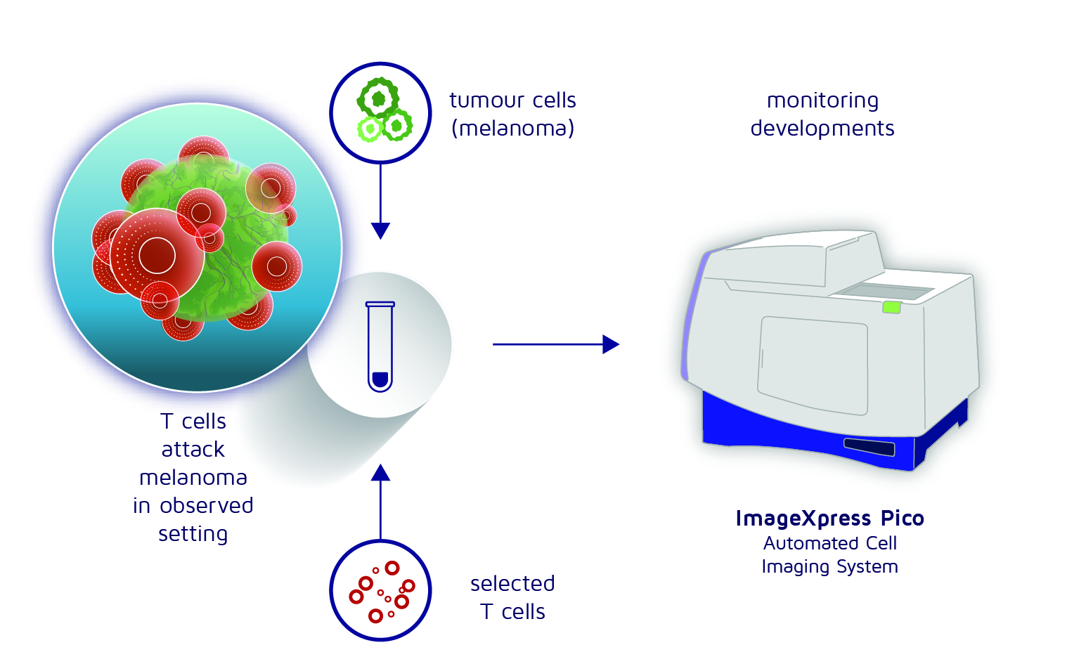
Figure 1: Procedure for testing T cells
We create spheroids of cancer cells, dyed green, to mimic a 3D tumor environment, and add red-colored T cells. We then place the test tube into an “automated cell imaging system”—a smart microscope that captures images regularly and automatically over several days. By tracking the disappearance of green cancer cells, we can measure the effectiveness of the T cells in destroying them.
But the cell population in a natural lymph node is not nearly as homogenous. And so, we acquire more insight if we analyze it piece by piece, using single-cell transcriptomic measurement. We are now as it were concerned with examining how many types of fruit there are, and how many of each we can identify. From this list, we acquire a far more precise picture.
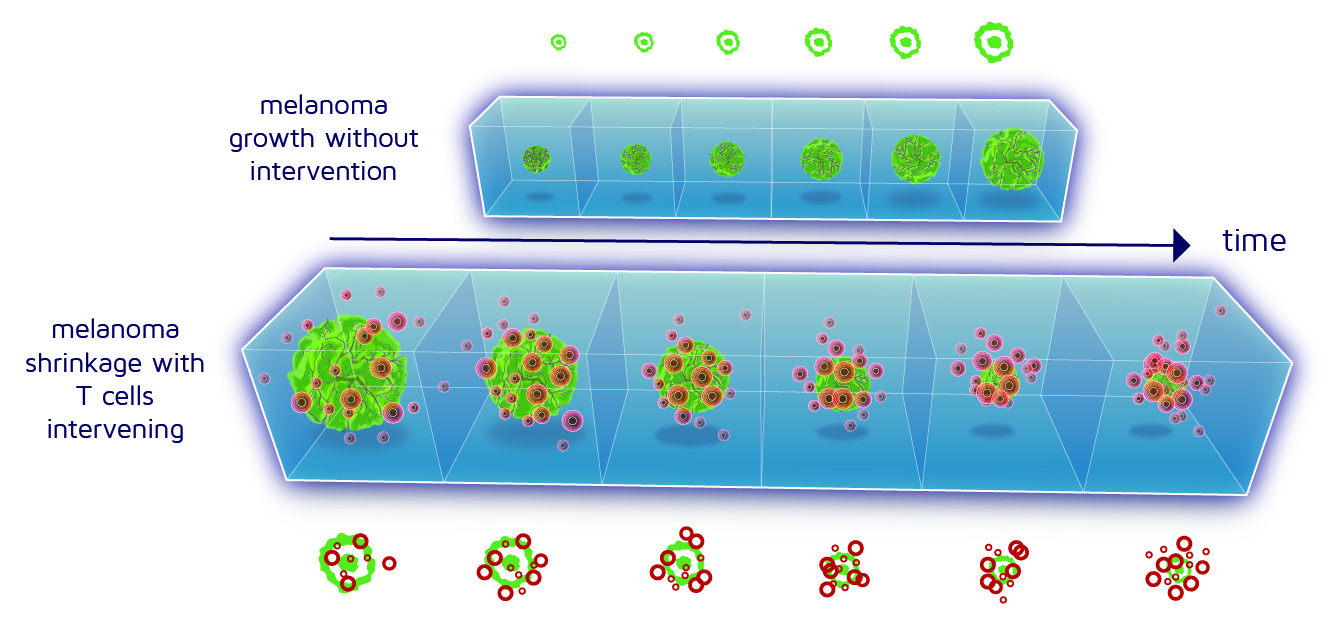
Figure 2: Development of a melanoma spheroid (green) without and with T cell intervention (red)
In vivo testing
To simulate a more challenging battlefield for the T cells, we add other cells found in actual cancers, such as macrophages or neutrophils, which can inhibit T cell function. This allows us to test whether, under these challenging conditions, the T cells truly become super soldiers after their training in our “artificial immune niche.”
Finally, we test the T cells’ behavior in real battle conditions. For mouse T cells, the battlefield will be mice carrying tumors, where the T cells must prove their combat effectiveness. This will be explained further below. For human T cells, we simulate the battlefield in laboratory test tubes.
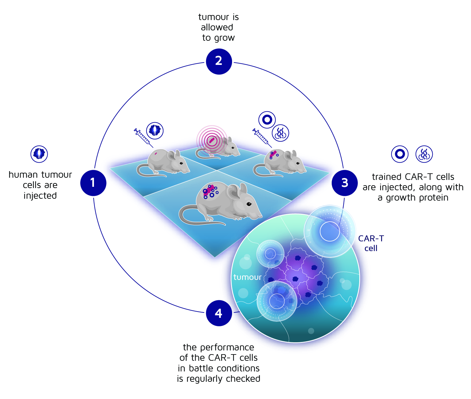
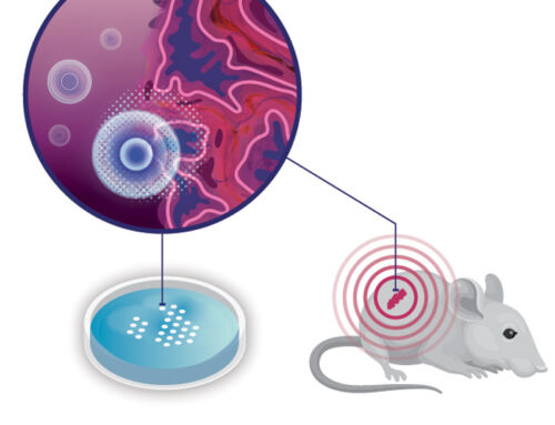
Figure 3: Procedure for in vivo testing
To test how well human T cells can fight tumors, scientists use special mice that lack a functioning immune system, allowing them to accept human cells without issues. First, human tumor cells are injected under the mice’s skin, where they grow into tumors. The mice are then given the “educated” CAR-T cells. To support the growth and activity of the CAR-T cells, the mice receive a protein that helps the T cells multiply, and scientists regularly measure the tumor size to see if the treatment is working. This sequence is depicted in figure 3.
By utilizing these methods, we acquire a relatively accurate picture about the efficacy of various types of CAR T cells. In turn, these insight helps us to select the best cells to fight cancer tumors.